Skip to main content Skip to navigation

Molecular biomarkers of canine and feline primary osteosarcoma

Osteosarcoma is an aggressive tumour in humans and in companion animals. The survival rate is low in humans and canines due to the high metastatic potential of the malignant bone tumour. Using proteomics approach (2D-gelelectrophoresis, mass spectrometry (MS), immunohistochemistry, zymography), gene expression profiling and unbiased RNA analysis on canine tumour cell lines and ex vivo canine and feline osteosarcoma primary cells we hope to identify specific biomarkers of osteosarcoma and improve the understanding of the tumour biology, in particular of the factors involved in metastasizing. Several project parts (see below) were developed since 2011. This project is worked on by the PhD students Mag. Florian Meyer and Dipl-Ing.(FH) Christiane Gebhard MSc, at the host Institute of Anatomy, Histology and Embryology (PI A. Univ. Prof. Ingrid Walter) in cooperation with VetCore (A. Univ. Prof. Steinborn, A. Univ. Prof. Razzazi), Oncology (Priv. Doz. Dr. Wolfesberger), Institute of Medical Chemistry (Ing. Ingrid Miller), Institute of Pathology (Dr. Andrea Fuchs-Baumgartinger), Univ. Prof. Obermayer-Pietsch (Meduni Graz).

Comparative histology of canine and feline osteosarcomas
The enormous heterogeneity of osteosarcomas influences all methodological approaches in osteosarcoma research and hampers interpretation of results. Therefore, a detailed morphological analysis and description of the morphology of each individual tumour is a prerequisite. Every incoming tumour is typed by histopathology, immunohistochemistry (metastasizing markers, angiogenesis markers), proliferation status and nature and extent of extracellular matrix.

2-dimensional and 3-dimensional cell culture of cell lines and primary canine and feline osteosarcoma cells
Osteosarcoma cell cultures were included in the study as they are relevant for experimental in vitro applications including pharmacological tests. Besides cultivating commercially available tumour cell lines, our approach is to obtain primary tumour cells from incoming clinical samples. These primary cells are characterized by bone-specific tumour markers. Furthermore, it is important to cultivate osteosarcoma cells in a 2D and 3D manner as expression patterns might differ significantly accompanied by altered tumour cell behaviour such as response to drug treatment. Therefore, we compare the morphology (on the light- and electron microscopic level) and the proteome pattern by immunohistochemistry, 2D electrophoresis , DIGE and MALDI-TOF/TOF.

Do Erythropoetin (Epo) and Erythropoetin-receptor (EpoR) have a role in canine or feline osteosarcoma progression?
Clinical data show that Epo does have a stimulatory effect on several tumour types. Beside the systemic Epo influence as originating from the kidney or therapeutically administered Epo, also tumour cells might be a source of Epo secretion, stimulating proliferation and angiogenesis. We attempt to find out if canine or feline osteosarcoma cells are capable of Epo or EpoR expression by means of PCR, immunohistochemistry and functional in vitro tests.

Activity of matrix metalloproteinases 2 and 9 in canine and feline osteosarcomas
MMP´s are important degrading factors in physiological and pathological processes such as metastasation. Due to the different metastasizing behaviour in feline compared to canine osteosarcomas we hypothesized that this difference might be reflected by MMP2 or MMP9 activity. Presence and activity of MMP2 and MMP9 are determined in osteosarcoma cell cultures and clinical samples by zymography and immunohistochemistry. A potential association with the tumour type is examined.

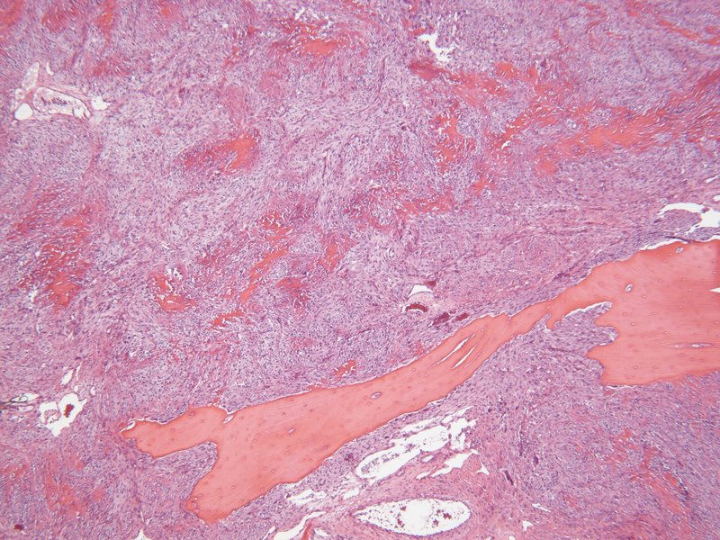
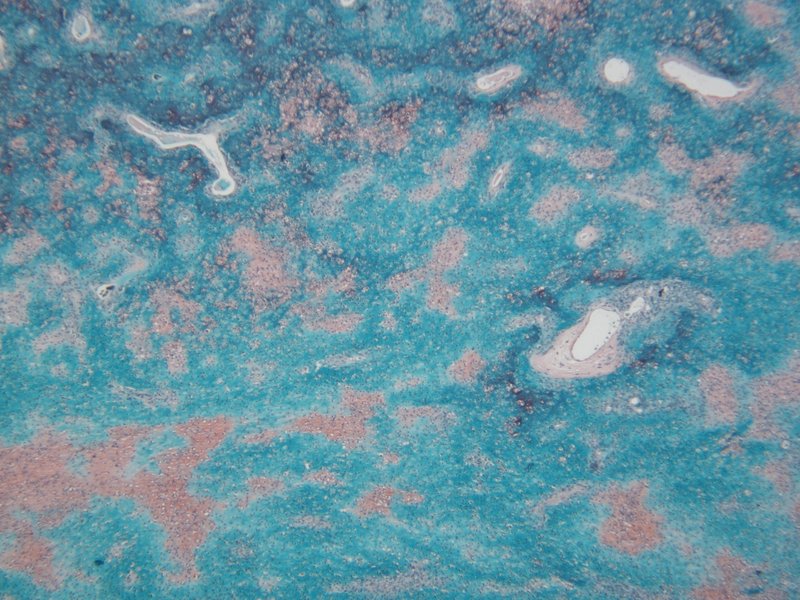
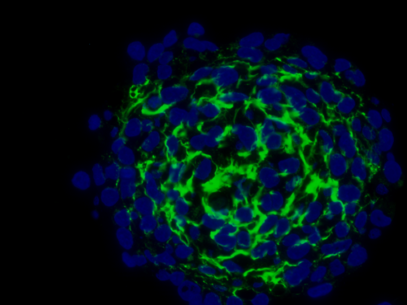
c-Kit (CD117) and kit ligand (stem cell factor) expression in canine and feline osteosarcomas
Tyrosine kinase inhibitors are modern pharmaceutical drugs which are able to block important signal transduction cascades essential for cancer cell growth. A potential candidate for a therapeutic application is the tyrosine kinase CD117 (c-kit, stem cell factor receptor), as this receptor is often overexpressed on tumour cells. CD117 positive cells showed high metastatic and drug-resistant properties in human osteosarcoma cells. In our study the presence and distribution of CD117 and its ligand (SCF) is determined on cultured osteosarcoma cellsand clinical tumour samples of dogs and cats by immunohistochemistry and western blot on the protein level and PCR on the mRNA level.  Knowing about the presence or the absence of potential targets in osteosarcoma  samples of companion animals will help in future to use targeted therapy in a specific individualized therapeutic approach in veterinary medicine as well.

Glycosylation patterns of osteosarcoma cells by lectin histochemstry
Cell surface carbohydrate changes may be responsible for the metastatic potential of malignant cells. Therefore, a differentiation between the various subtypes of osteosarcoma based on lectin-binding patterns might be possible. Therefore, we apply lectin histochemistry to analyse a panel of glycoconjugates and demonstrate the distribution of ascertained specific sugar residues within the different types of heterogeneous canine and feline osteosarcoma tumors.

Strategy for reference genes in osteosarcoma research
The analysis of transcript expression by Reverse transcription quantitative PCR (RT-qPCR) requires appropriate data normalization compensating for biological and technical fluctuations. The accuracy of RT-qPCR data was shown to improve from a normalization strategy specifically adjusted to the biological context of the experimental study, for example a specific type of tissue, a physiological or disease state, etc. Here we apply an educated guesswork-free approach selecting reference sequences  from high-throughput RNA expression data for the context of canine osteosarcoma. The geometric averaging of multiple internal control sequences specifically selected for the experimental context will allow more accurate normalization of RT-qPCR data.

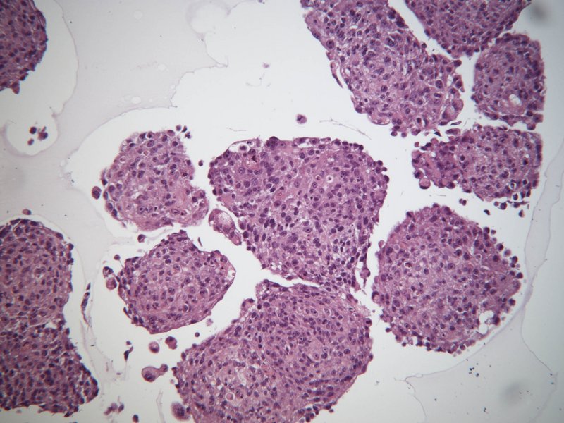
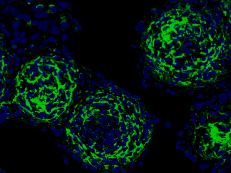
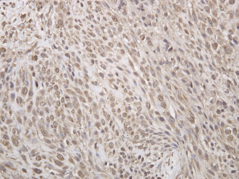
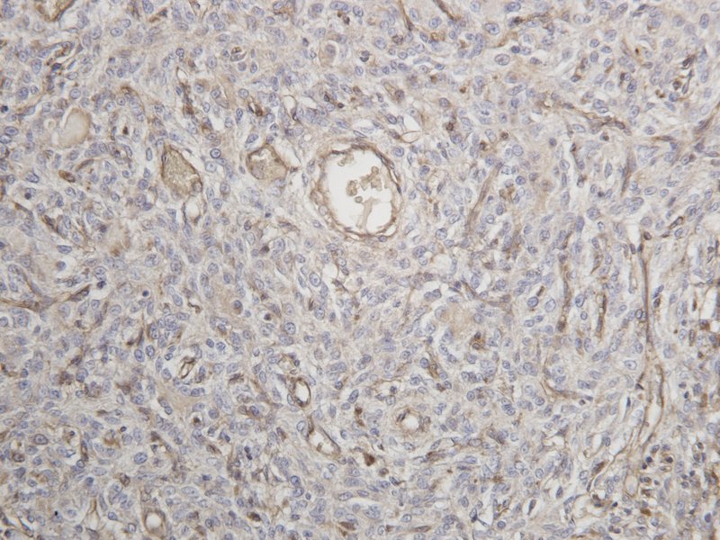
Osteosarcoma associated macrophages
Tumour associated macrophages are critical factors for tumour progression and metastases by cytokine release. They might have a supporting or inhibiting function on different types of tumour cells depending on their activation status. Data regarding osteoclasts are conflicting as it has been reported that their ablation leads to an increase or reduction of tumour burden. We attempt to analyse the different macrophage populations within canine and feline osteosarcomas (including giant cells and osteoclasts) by means of immunohistochemistry.